
Auricular hematoma in dogs and cats – treatment in Gdańsk
The ear suddenly swelled, is soft, warm, and looks like a pillow? This is most often a hematoma. It’s better not to delay the visit: quick treatment prevents permanent deformation of the auricle, and outpatient puncture of the hematoma immediately brings relief to the animal.

What is an auricular hematoma?
It is a collection of blood between the skin and the cartilage of the ear. It occurs when the pet shakes its head vigorously or scratches its ear, causing small blood vessels to rupture. The most common cause is itching and ear inflammation.
Treatment has two goals: to remove the blood from the ear and to treat the underlying cause. Without treating the cause, the hematoma often returns, and if left untreated, it leaves a thickened, wrinkled ear.
What happens to the ear
See how the ear changes without treatment.
Illustrative diagram. Created by AI.
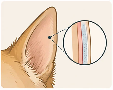
Healthy auricle
The skin tightly adheres on both sides to the thin cartilage. The ear is flat, light, and flexible.
What will you notice
The ear has an even thickness, it is neither warm nor painful.
Illustrative diagram. Created by AI.
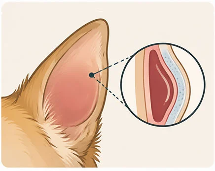
Ear hematoma
Blood collects between the skin and cartilage. The ear becomes thick like a pillow, heavy, and warm. This is a good time for treatment, as the ear has the best chance of returning to its former appearance.
What will you notice
Sudden, soft swelling of the ear, usually from the inside. The pet shakes its head and tilts it to the side.
Illustrative diagram. Created by AI.
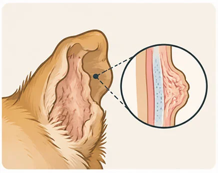
Deformed ear
The blood gradually absorbs, but scar tissue forms in its place, which shrinks and wrinkles the ear. Such a change cannot be fully reversed.
What will you notice
A hard, thick, wrinkled ear that may hinder ventilation of the ear canal.
Hematoma is a consequence. The most important thing is to find the cause
A hematoma is almost always the result of something that itches or hurts. Therefore, with every hematoma, we also examine the ear canal.
→
1. Itching or pain in the ear
Ear inflammation, allergy, ear mites, foreign body.
→
2. Shaking the head
The pet energetically shakes its ears or scratches them with its paw.
→
3. Small vessel bursts
Blood collects between the skin and the ear cartilage.
4. Hematoma
The ear swells, feels heavy, and continues to itch, so the pet shakes it even harder.
In dogs
Ear itching is most often caused by allergies and external ear inflammation.
Dogs with floppy ears, such as golden and labrador retrievers, cocker spaniels, or basset hounds, are more frequently affected.
In cats
Hematomas occur less frequently in cats than in dogs. They are most often caused by ear mites.
How we treat a hematoma – comparison of methods
There is no one method for all. We choose it together with you after examining the ear.
Puncture with a needle (blood withdrawal)
The doctor withdraws blood from the ear with a needle. Sometimes an anti-inflammatory medication is also administered inside the hematoma or orally.
Advantages
The least invasive method, without skin incision. Good appearance of the ear if the hematoma does not recur.
Limitations
A hematoma after the puncture is more likely to return. Sometimes it needs to be repeated.
When we choose it
For small, fresh hematomas or when general anesthesia poses a significant risk to the pet.
Drainage
We leave a thin drain in the hematoma through which the fluid drains, and the skin adheres to the cartilage. The drain and stitches are removed after about 3 weeks.
Advantages
Blood and fluid drain continuously, without a large incision.
Limitations
Requires monitoring of the drain for about 3 weeks and follow-up visits. The pet cannot pull it out.
When we choose it
For moderate hematomas and in patients where we want to avoid a larger procedure.
Surgical procedure
We make an incision on the inner side of the ear, remove blood and clots, and then attach the skin to the cartilage with stitches to avoid leaving a free space. We often also place a drain. Stitches and drain are removed after about 3 weeks.
Advantages
Lowest risk of recurrence. Good effect even with large and recurring hematomas.
Limitations
Requires general anesthesia. The ear may be slightly thickened after the procedure.
When we choose it
For large hematomas, recurrences, or when puncture did not help.
CO₂ Laser Treatment
Using a laser, we create several small, round holes in the skin on the inner side of the ear. Through these, blood drains, and the skin fuses with the cartilage.
Advantages
Less bleeding during the procedure. The openings allow drainage without a long incision.
Limitations
Requires general anesthesia and adherence to recommendations until the openings heal.
When we choose it
As an alternative to the classic procedure, especially in cases of recurring hematomas.
How the treatment proceeds – step by step
At each stage, you know what is happening and what comes next.
1
Ear examination
We examine the ear canal (otoscopy) and analyze the discharge under a microscope to find the cause of itching.
2
Method selection
We discuss options with you: puncture, drainage, procedure, or laser. We consider the size of the hematoma and whether it recurs.
3
Procedure
We remove blood and clots, and attach the skin to the cartilage with stitches, often with a drain. The pet usually goes home the same day.
4
Treatment of the cause and monitoring
We treat ear inflammation or allergy. After about 3 weeks, we remove the stitches and drain. Treating the cause is key to preventing the hematoma from returning.
What you should know before treatment
- The needle puncture itself is less invasive, but the hematoma is more likely to return after it. Surgical treatment is less likely to result in a recurrence.
- The ear may be slightly thickened or wrinkled after treatment, especially if the hematoma was large or lasted a long time.
- If the cause of itching is not treated, the hematoma may reappear, also in the other ear.
Care after hematoma treatment
Protective collar
Protects the ear from scratching with the paw. Wear it until the doctor allows you to remove it.
Dressing or ear protector
If we have applied it, it limits ear flapping. Make sure it stays dry and does not apply pressure.
Ear drops and medications
Administer them exactly as directed, even when the ear looks good.
Removal of stitches and drain after about 3 weeks
Until then, do not touch the drain. At follow-up visits, we check the healing and whether the ear inflammation is subsiding.
Call immediately if:
- the ear has clearly swollen again,
- pus is leaking from the wound or it has an unpleasant smell,
- the drain has fallen out, the dressing has slipped, soaked, or is applying pressure,
- the pet tilts its head, loses balance, or is not eating.
Our entire team of surgeons operates on ear hematomas



Questions about ear hematoma
Is your ear swelling and you don’t know what to do? Call us, we will help assess how urgent the visit is.
FAQ – questions and answers
Based on research
Hewitt J., Bajwa J. (2020), Canadian Veterinary Journal
A review of the causes and treatment methods for aural haematomas in dogs and cats, from puncture to surgical procedures and CO₂ laser. – Can Vet J 2020;61(3):313–315 →
Hall J. et al. (2016), Journal of Small Animal Practice
A survey of veterinarians in the UK: how they treat haematomas and which method they assign the least risk of recurrence. – doi: 10.1111/jsap.12524 →
Don’t wait for the ear to become deformed
The sooner we start treatment, the better the appearance of the ear and the lower the risk of recurrence.