
Ear canal surgeries for dogs and cats in Gdańsk
Does ear inflammation return despite drops, antibiotics, and flushing?
The ear canal is narrowed, painful, and constantly dirty?
When conservative treatment no longer helps, ear canal surgery can permanently free your pet from pain and itching.
When is ear canal surgery performed?
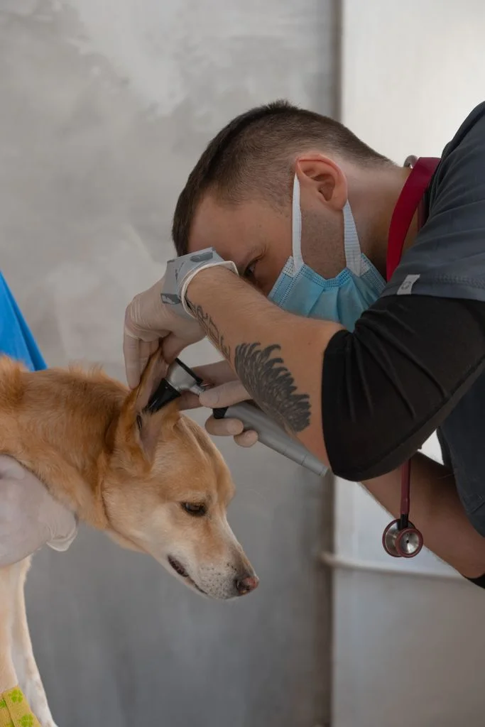
Surgery is considered for chronic ear inflammation that does not respond to treatment, for a narrowed or calcified canal, and for polyps and tumors. The indication is determined by the veterinarian after examination and imaging diagnostics, such as otoscopy and computed tomography.
The most common procedures are vertical canal ablation, polyp removal in cats, and total ear canal ablation (TECA). TECA is a definitive surgery when other treatment methods are ineffective.
The decision about surgery is made by the veterinarian – after examination and diagnostics
Based on symptoms at home, it is not possible to assess whether the ear requires surgery. The decision for surgical treatment is made only after clinical examination and additional tests, including imaging diagnostics.
What to bring for the ear examination
- ear inflammation returns despite treatment
- unpleasant smell and discharge from the ear despite drops
- the pet does not allow touching the ear, scratches it, or shakes its head
- hard lump at the base of the ear
- nodule or growth in the ear canal
What we check before making a decision
- clinical examination and skin assessment
- otoscopic examination of the ear canal, if necessary under anesthesia
- cytology and culture of discharge with antibiogram
- computed tomography (CT) of the head – assessment of the ear canal and middle ear
- blood tests before anesthesia
Urgent
Is your pet tilting its head or losing balance?
This may indicate that the inflammation has affected the middle or inner ear.
Contact us today.
Types of ear canal operations
Choose a procedure. We have marked which part of the ear it concerns on the diagram.
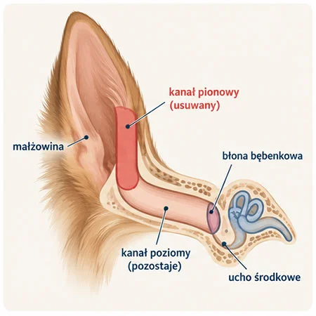
Illustrative diagram. Created by AI.
Vertical ear canal ablation
We remove the entire vertical segment of the ear canal, while the horizontal canal remains and opens directly to the outside.
When we perform it
When changes, such as growths or tumors, involve the vertical canal, while the horizontal canal and middle ear are healthy.
What is worth knowing
Hearing is usually preserved. A thorough assessment is required to ensure that the horizontal canal is not affected.
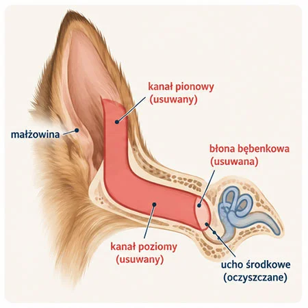
Illustrative diagram. Created by AI.
Total Ear Canal Ablation (TECA)
We remove the entire diseased ear canal and clean the middle ear (tympanic bulla osteotomy). The pinna remains, so the dog looks as it did before.
When do we perform it
This is a definitive surgery. We perform it when changes in the canal are so advanced (narrowing, calcification, involvement of the middle ear) that other treatment methods will be ineffective. Also for certain tumors of the ear canal.
What is worth knowing
This is the most effective surgery for chronic inflammation. The dog hears worse on the operated side, and the risk of irritation of the facial nerve is discussed before the procedure.
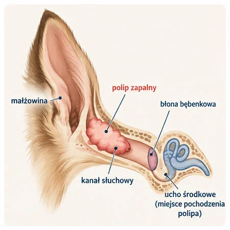
Illustrative diagram. Created by AI.
Removal of Ear Polyps in Cats
Inflammatory polyps grow from the middle ear and can invade the ear canal or throat. We remove them through the ear canal, and in cases of recurrence or large polyps, we also clean the middle ear.
When do we perform it
In cats, most often young ones, with discharge from the ear, head tilting, snoring, or a visible lump in the ear.
What is worth knowing
After the removal of the polyp, it may regrow. Cleaning the middle ear reduces this risk, but it is a more extensive procedure.
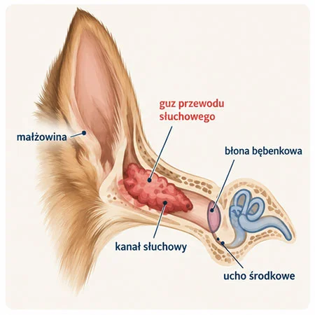
Illustrative diagram. Created by AI.
Removal of Tumors in the Ear Canal
Tumors in the ear, such as ceruminous gland adenomas, can block the canal and sustain inflammation. The extent of the surgery depends on the size and type of the lesion.
When do we perform it
When there is a growth in the ear, the ear is bleeding, or the inflammation is only on one side and does not subside.
What is worth knowing
The removed lesion is always examined histopathologically. In the case of malignant changes, TECA is usually necessary.
How the treatment proceeds – step by step
Ear surgery is a big step, so before making a decision, we carefully check what is happening in the ear canal and in the middle ear.
1
Diagnostics
Clinical examination, otoscopic examination, computed tomography (CT), cytology and culture of the discharge with an antibiogram, and blood tests before anesthesia.
2
Doctor’s decision
Based on the results, the doctor assesses whether surgery is needed and what type. We discuss with you the risks and how your pet’s hearing will change.
3
Surgery
Under general anesthesia with full monitoring and strong pain management. The removed tissue is sent for histopathological examination.
4
Recovery
The pet remains under 24-hour care at the clinic for as long as needed. At home, you continue treatment according to the written instructions.
What is worth knowing before ear surgery
- The type of surgery depends on the severity of the changes visible in the examinations. TECA is a definitive surgery – we perform it when other treatment methods are ineffective.
- After TECA, the dog hears worse on the operated side. With advanced disease, hearing is usually already significantly impaired, so owners often do not notice the difference.
- In surgeries around the middle ear, there is a risk of irritation of the facial nerve, e.g., weaker blinking. It is usually temporary. We discuss this before the procedure.
- The surgery removes the diseased canal but does not treat allergies. The skin and ears need to be monitored further.
Post-ear surgery care
Protective collar
Protects the wound from scratching with the paw and rubbing against furniture. Keep it on until the doctor allows you to remove it.
Painkillers and antibiotics
Administer them exactly as directed. The antibiotic is selected based on the culture.
Harnesses instead of collars
The collar may press on the area of the wound. Walks only on a leash.
Follow-up visits
We check the healing of the wound and discuss the results of the histopathological examination.
Call immediately if:
- the ear has clearly swollen again,
- pus is leaking from the wound or it has an unpleasant smell,
- the drain has fallen out, the dressing has slipped, soaked, or is applying pressure,
- the pet tilts its head, loses balance, or is not eating.
Ear canal surgeries at Sosken are performed by


How we treat hematomas – comparison of methods
There is no one method for everyone. We choose it together with you after examining the ear.
Puncture with a needle (blood withdrawal)
The doctor drains blood from the ear with a needle. Sometimes an anti-inflammatory medication is also administered inside the hematoma or orally.
Advantages
The least invasive method, without cutting the skin. Good appearance of the ear if the hematoma does not recur.
Limitations
A hematoma after just a puncture is more likely to return. Sometimes it needs to be repeated.
When we choose it
For small, fresh hematomas or when general anesthesia poses a significant risk to the pet.
Drainage
We leave a thin drain in the hematoma through which fluid drains, and the skin adheres to the cartilage. The drain and stitches are removed after about 3 weeks.
Advantages
Blood and fluid drain continuously, without a large incision.
Limitations
Requires monitoring of the drain for about 3 weeks and follow-up visits. The pet cannot pull it out.
When we choose it
For medium hematomas and in patients where we want to avoid a larger procedure.
Surgical procedure
We cut the skin from the inner side of the ear, remove blood and clots, and then attach the skin to the cartilage with stitches to avoid leaving any free space. We often also place a drain. Stitches and drain are removed after about 3 weeks.
Advantages
The lowest risk of recurrence. Good results even with large and recurrent hematomas.
Limitations
Requires general anesthesia. The ear may be slightly thickened after the procedure.
When we choose it
In cases of large hematomas, recurrences, or when puncture did not help.
CO₂ Laser Treatment
Using a laser, we create several small, round holes in the skin on the inner side of the ear. Through these, blood drains out, and the skin heals to the cartilage.
Advantages
Less bleeding during the procedure. The holes allow drainage without a long incision.
Limitations
Requires general anesthesia and adherence to recommendations until the holes heal.
When we choose it
As an alternative to the classic procedure, especially in cases of recurrent hematomas.
Questions about ear surgery
Has your dog been struggling with ear inflammation for months? Schedule a consultation, and together we will assess whether surgery makes sense.
FAQ – questions and answers
Based on research
Coleman K.A., Smeak D.D. (2016), Veterinary Surgery
Complications after total ear canal ablation with bulla osteotomy (TECA-LBO) in dogs – surgery of one ear vs both ears in one procedure (79 ears). – doi: 10.1111/vsu.12505 →
Anderson D.M. et al. (2000), Veterinary Record
Treatment of inflammatory polyps in 37 cats – comparison of polyp removal via the ear canal or throat with bulla osteotomy. – doi: 10.1136/vr.147.24.684 →
Chronic ear pain does not have to last for years
Schedule a surgical consultation. We will assess the ear condition and present possible solutions.