
Salivary gland cyst in dogs and cats – treatment in Gdańsk
A soft, painless swelling under the jaw, on the neck, or under the tongue that grows slowly?
This may be a salivary gland cyst, which is saliva accumulating under the skin after damage to the gland or salivary duct. It will not go away on its own, but well-conducted surgical treatment offers very good prognosis.
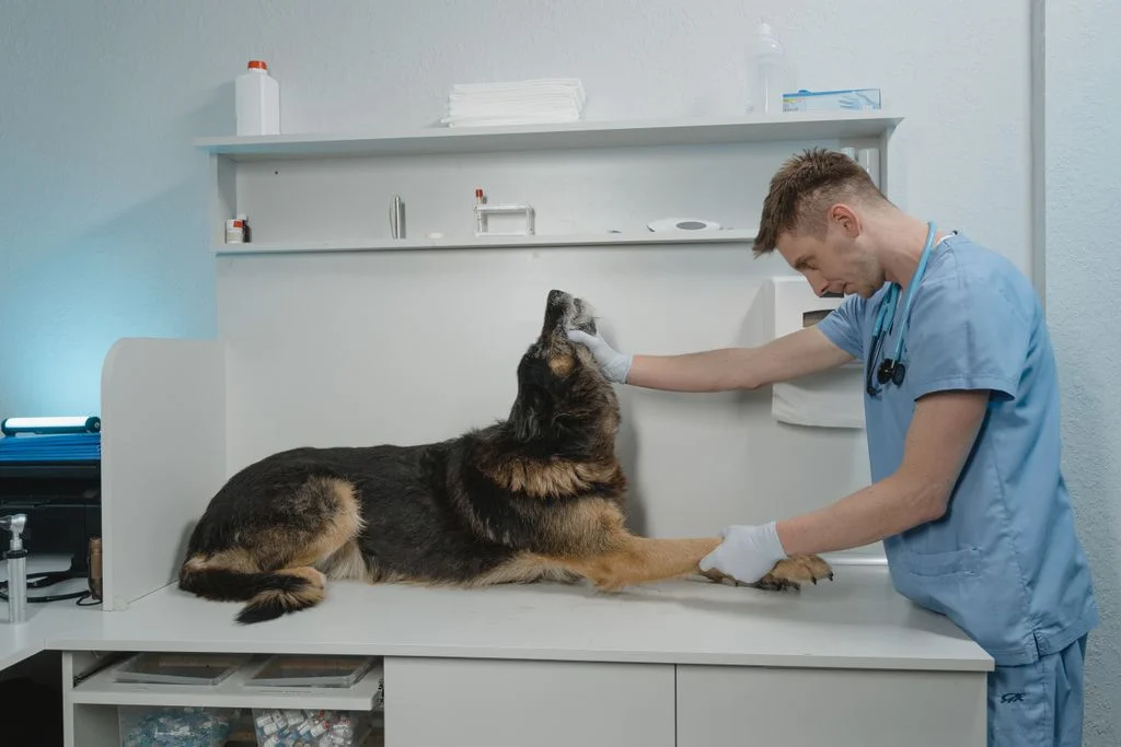
What is a salivary gland cyst?
It is an accumulation of saliva in the tissues around the damaged salivary gland or its duct. It most often originates from the sublingual gland and appears on the neck, under the jaw, or under the tongue.
Simply draining the fluid provides only temporary relief. The treatment of choice is the surgical removal of the diseased salivary glands on the cyst side. The method is determined by the doctor after examination.
Where can a salivary gland cyst appear?
Choose a location to see the symptoms and how urgently action needs to be taken.
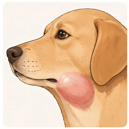
Illustrative diagram. Created by AI.
Cervical cyst
Saliva collects under the skin under the jaw or in the upper part of the neck. This is definitely the most common form of salivary gland cyst in dogs.
What will you notice
A soft, cool, painless swelling that grows slowly. The pet usually feels well.
How we treat
Removal of the submandibular and sublingual salivary glands on the side of the cyst.
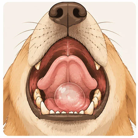
Illustrative diagram. Created by AI.
Sublingual cyst
A saliva-filled blister appears at the bottom of the mouth, next to the tongue. It often occurs together with a cyst in the neck.
What will you notice
Drooling, reluctance to eat, blood in saliva, bluish or pink blister under the tongue.
How we treat
Marsupialization of the cyst, often combined with the removal of the salivary glands.
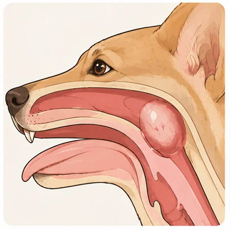
Illustrative diagram. Created by AI.
Pharyngeal cyst
Saliva collects in the wall of the throat and can narrow the airways. The swelling is often not visible from the outside.
What will you notice
Snoring, loud breathing, shortness of breath, difficulty swallowing, gag reflex.
How we treat
Urgent drainage of the cyst, followed by removal of the diseased salivary glands.
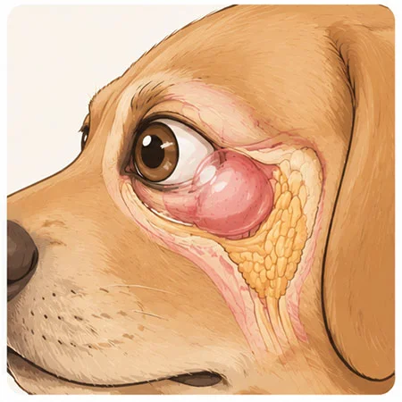
Schematic diagram. Created by AI.
Cheek salivary gland cyst
The cheek salivary gland is located under the eye. When a cyst forms in it, it can push the eyeball outward.
What will you notice
Swelling under the eye, protrusion of the eyeball, visible third eyelid, pain when opening the mouth.
How we treat it
Removal of the cheek salivary gland after thorough imaging diagnostics.
Is it definitely a salivary gland cyst?
Clinical examination
Assessment of swelling, oral cavity, and area under the tongue.
Fine needle aspiration
Thick, viscous, yellowish or blood-tinged fluid indicates saliva. We examine it under a microscope.
Neck ultrasound
Shows the size and extent of the cyst and helps differentiate it from other lesions.
Computed tomography (CT)
If necessary, e.g., in case of recurrences or when it is difficult to determine from which side the cyst originates.
Urgent
Does your pet have difficulty breathing or swallowing?
A cyst in the throat can narrow the airways. This is an emergency – do not wait for a scheduled appointment.
Why simply draining saliva is not enough
The damaged salivary gland continues to produce saliva, so after just puncturing the cyst, it usually returns. A lasting effect is only achieved by removing its source.
Cyst puncture
Draining saliva with a needle. It provides relief and is part of the diagnostics, but does not treat the cause, so the cyst usually fills up again.
Removal of salivary glands
We remove the submandibular and sublingual glands on the side of the cyst and empty the cyst itself. This removes the source of saliva and provides a lasting effect.
Marsupialization
For a cyst under the tongue, we open it and suture the edges so that saliva can freely flow into the oral cavity. We often combine this with the removal of salivary glands.
How treatment proceeds – step by step
Before the surgery, we precisely determine which salivary gland is the source of the cyst.
1
Diagnostics
Examination, puncture with fluid assessment, ultrasound, and if necessary, CT. Blood tests before anesthesia.
2
Doctor’s decision
The doctor determines which salivary gland is the source of the problem and discusses the treatment plan with you.
3
Surgery
Under general anesthesia, we remove the diseased salivary glands and empty the cyst. The decision to place a drain is made by the operating surgeon. The removed tissue is examined histopathologically.
4
Return and follow-up
Written recommendations and a follow-up visit. The pet is under 24-hour care at the clinic for as long as necessary.
What to know before the surgery
- A dog or cat without removed salivary glands eats normally and does not have dry mouth. The remaining salivary glands produce enough saliva.
- After the surgery, swelling with fluid may appear at the cyst site for a few days. It usually resolves on its own, but show it to the doctor during the follow-up.
Post-surgery care for salivary glands
Collar or protective clothing
Protects the wound on the neck from scratching with the paw. Wear it until the doctor allows you to remove it.
Harness instead of a collar
A collar puts pressure on the wound on the neck. Walks only on a leash and in a harness.
Soft food
For a few days, provide soft food, especially after the procedure under the tongue. No chews or hard toys.
Medications and follow-up visit
Administer medications as directed. If the surgeon placed a drain, do not touch it – we will remove it during the follow-up. We will also discuss the results of the histopathological examination.
Call immediately if:
- the pet has difficulty breathing or swallowing,
- swelling on the neck is rapidly increasing,
- the gland is bleeding, pus is oozing from it or it has ruptured, or the drain has fallen out,
- it is not eating, is apathetic or has a fever.
Salivary gland surgeries at Sosken are performed by


Questions about salivary gland cysts
Have you noticed swelling on your pet’s neck or under its tongue? Schedule an examination, and we will explain what it is.
FAQ – questions and answers
Based on research
Ritter M.J. et al. (2006), New Zealand Veterinary Journal
Mandibular and sublingual salivary gland cysts in dogs – analysis of 41 cases treated surgically. – Full text →
Cinti F. et al. (2021), Veterinary Surgery
Comparison of complications after removal of the mandibular and sublingual salivary glands via ventral and lateral approaches in dogs with salivary cysts. – doi: 10.1111/vsu.13601 →
Swelling on the neck? Let’s check what it is
Schedule an examination. The doctor will make a diagnosis and propose treatment.