
Oncological surgery for dogs and cats in Gdańsk
A cancer diagnosis for your pet is a difficult moment. We will help you understand what comes next. Before we plan the surgery, we check what type of cancer we are dealing with and whether it has spread. The surgeon plans the operation based on the test results, and you receive clear information about the purpose of the surgery, the risks, and other options.
Content author: veterinarian Małgorzata Szczepańska
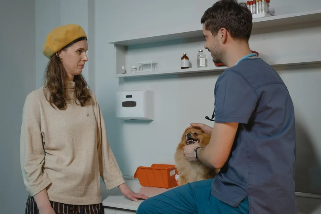
What is oncological surgery for dogs and cats?
It is the surgical treatment of tumors. The surgeon removes the tumor along with a margin of healthy tissue, and the removed tissue is always sent for histopathological examination, which confirms the diagnosis and assesses the margins.
For many solid tumors, surgery is the most effective treatment method. For some cancers, additional treatment, such as chemotherapy, is needed after surgery – this is indicated by the results of the histopathological examination.
Four questions we answer before surgery
1
What kind of lump is it?
Cytology from a fine needle biopsy or a sample for histopathological examination.
2
Has it spread?
Chest X-ray, abdominal ultrasound, and lymph node assessment, and if necessary, a computed tomography (CT).
3
Will the pet tolerate the procedure?
Blood tests and assessment of overall health, and if necessary, heart examination.
4
What is the purpose of the surgery?
Cure, reduce the tumor, or improve comfort – we determine this with you before the procedure.
Why don’t we operate “right away”
The type of cancer determines how extensively it needs to be removed and whether surgery is even the best choice.
The first surgery gives the greatest chance of completely removing the tumor, which is why it is worth planning it well.
This does not mean a long wait. We try to carry out diagnostics as quickly as possible.
What purpose can surgery have
Not every oncological surgery has the same purpose.
We talk about this openly so that you know what to expect.
Radical operation
We remove the entire tumor with an appropriate margin of healthy tissue. The goal is to cure or achieve long-term control of the disease.
When we propose it
When the tumor is confined to one location, and tests do not show metastases.
Tumor-reducing operation
We remove as much of the tumor as possible when it cannot be completely removed. After such an operation, further treatment, such as chemotherapy, is usually needed.
When we propose it
When the tumor invades important structures, and reducing its mass will improve the effectiveness of further treatment.
Comfort-improving operation
The goal is not to cure, but to improve the quality of life: removal of a painful, bleeding, or ulcerated tumor or restoring, for example, the ability to pass stool freely.
When do we recommend it
When the disease is advanced, but the procedure will realistically improve the pet’s quality of life.
Types of tumors we most often treat surgically
Select to see what to pay attention to and how we proceed. Diagnosis and treatment plan are always established by the veterinarian after examinations.
Milk line tumors
One of the most common tumors in unspayed females
What you should know
In females, about half of milk line tumors are malignant. In cats, the vast majority of such tumors are malignant, so any lump near the nipple in a cat requires urgent diagnostics.
What to pay attention to
Hard or soft lumps along the mammary chain, sometimes several at once. They may grow, become red, or ulcerate.
How we proceed
Before the surgery, we perform a chest X-ray and an abdominal ultrasound. The scope of the procedure – from the removal of a single gland to the removal of the entire mammary chain – is determined by the surgeon. For female cats, we usually recommend removing the entire chain.
Spleen tumors
Often detected incidentally during an ultrasound examination
What is worth knowing
Spleen tumors can be benign or malignant, e.g., hemangiosarcoma. They cannot be distinguished without histopathological examination.
What to pay attention to
Enlarged abdomen, weakness, pale gums. A rupture of the tumor causes bleeding into the abdominal cavity – then call and come immediately.
How we proceed
We remove the entire spleen (splenectomy) and send it for histopathology. Before the procedure, we look for changes in other organs. After the surgery, the pet remains under 24-hour care at the clinic.
Anal area tumors
Perianal adenomas and tumors of the anal sac glands
What is worth knowing
Perianal adenomas are usually benign and mainly affect unneutered males. Tumors of the anal sac glands are more often malignant and can metastasize to lymph nodes in the pelvis.
What to pay attention to
A lump near the anus, bleeding, difficulty in passing stool. Sometimes increased thirst when the lump raises calcium levels in the blood.
How we proceed
We remove the lump and send it for histopathology. In unneutered males with adenoma, we usually also perform castration. For anal sac tumors, we assess the lymph nodes via ultrasound before surgery.
Testicular tumors
More common in dogs with undescended testicles (cryptorchidism)
What is worth knowing
Most testicular tumors in dogs can be treated with castration. Some of them produce hormones and cause skin changes or enlargement of nipples in males.
What to pay attention to
Enlarged or hard testicle, varying testicle sizes, symmetrical bald patches, enlarged nipples.
How we proceed
Castration with histopathological examination. In cases of internal tumors, we also remove the testicle from the abdominal cavity or from the groin.
Skin and soft tissue tumors
Including tumors from mast cells and soft tissue sarcomas
What is worth knowing
The most common cancers in dogs. Some of them are malignant, and it is not possible to assess this by appearance.
What to pay attention to
A lump that grows, changes appearance, bleeds, or persists for more than a month.
How we proceed
First cytology, then a procedure with a margin tailored to the type of tumor – scalpel or CO₂ laser. More on the page about skin tumor removal.
Treatment step by step
At every stage, you know what is happening, and you can call with questions.
1
Consultation
Examination, discussion of symptoms and previous results, diagnostic plan.
2
Diagnostics
Cytology or biopsy and imaging studies that show the extent of the disease.
3
Surgery
Under anesthesia with full monitoring and pain management. If necessary, blood supply from our blood bank.
4
Histopathology
Final diagnosis, degree of malignancy, and assessment of whether the tumor was completely removed.
5
Further plan
We discuss the results and further steps, including any additional treatment. We set control dates.
What to know before making a decision
- Not every cancer can be cured by surgery. Sometimes its goal is to prolong life in good comfort – and we speak about this directly.
- A histopathological examination may show that the tumor has not been completely removed. Then we discuss a second procedure or additional treatment.
- Sometimes surgery is not the best choice for your pet. In that case, we propose other treatment options and palliative care.
- You always make the decision – after discussing the goals, risks, and quality of life for your pet.
Post-operative care for oncology
Collar or outfit
Protects the wound from licking until healed and stitches are removed.
Pain management
Administer medications as directed. Your pet should not feel pain – if you see that they are suffering, call.
Limited movement
Walks on a leash, no jumping or running until the wound is healed.
Results and controls
We discuss the histopathology results and set controls, e.g., imaging studies every few months.
Call immediately if:
- the wound is bleeding, oozing pus, or has opened up,
- your pet is very weak, pale, or having difficulty breathing,
- the abdomen is swelling after abdominal surgery,
- your pet is not eating, vomiting, or has a fever.
Who will take care of your pet



Questions about surgical treatment of tumors
Do you have test results and don’t know what to do next?
Schedule a consultation – we will calmly discuss all options.
FAQ – questions and answers
Based on research
Morris J. (2013), Journal of Feline Medicine and Surgery
Mammary tumors in female cats: significantly more malignant than in bitches, and tumors smaller than 3 cm are associated with better prognosis. Early, appropriately extensive surgery saves lives. – doi: 10.1177/1098612X13483237 →
Sorenmo K.U. et al. (2009), Veterinary and Comparative Oncology
Mammary tumors in bitches as a continuum from benign to malignant changes – an argument for early diagnosis and removal of tumors. – doi: 10.1111/j.1476-5829.2009.00184.x →
Liptak J.M. (2020), Veterinary and Comparative Oncology
Evaluation of histological margins after tumor removal – why histopathological examination of margins is crucial for further treatment planning. – doi: 10.1111/vco.12555 →
Let’s talk about the treatment plan
Bring your previous test results. We will explain the options and help you make a decision.