
Cherry eye in dogs – prolapse of the third eyelid gland. Treatment in Gdańsk
A red, smooth ball in the inner corner of the eye?
This is most often a prolapsed third eyelid gland, commonly referred to as “cherry eye.”
It is not an emergency condition, but it is better not to delay: the sooner we restore the gland to its place, the greater the chance that it will continue to produce tears properly.
What is cherry eye?
It is the prolapse of the third eyelid gland from its natural position. The gland protrudes in the inner corner of the eye as a red, swollen ball. It most commonly affects young dogs of predisposed breeds.
The treatment of choice is surgical repositioning of the gland using a pocket technique. The gland is not removed because it produces a large portion of tears. The pet usually returns home the same day.
What cherry eye looks like
Compare a healthy eye, an eye with a prolapsed gland, and an eye after surgery.
Illustrative diagram. Created by AI.
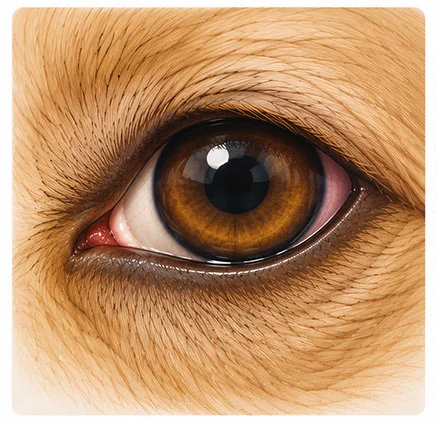
Normal third eyelid
The third eyelid is a fold in the inner corner of the eye. The gland hidden beneath it produces a large portion of tears. Normally, the gland is not visible.
What will you notice
In the corner of the eye, you can see a narrow, pink edge of the third eyelid.
Illustrative diagram. Created by AI.
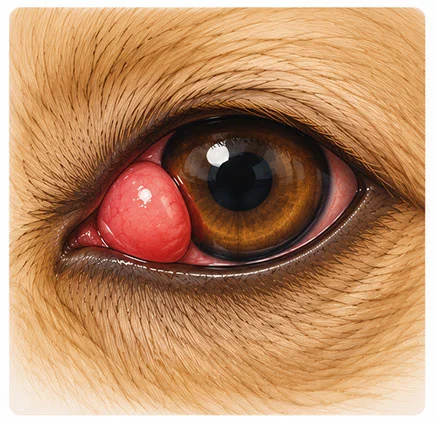
Cherry eye – cherry eye
The ligament that holds the gland in place is too weak. The gland protrudes, swells, and dries out. The longer it is outside, the more irritated it becomes.
What will you notice
A smooth, red ball in the inner corner of the eye, tearing, rubbing the eye with a paw.
Illustrative diagram. Created by AI.
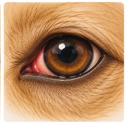
Gland back in place
After the procedure, the gland is back under the third eyelid and continues to produce tears. For a few days, the corner of the eye may be slightly swollen.
What will you notice
The eye looks as it did before the gland fell out, after a few days without swelling.
40–60%
the watery part of tears is produced by the third eyelid gland
Why we do not remove the gland
Removing the protruding gland seems like the simplest solution, but it significantly increases the risk of dry eye (dry keratitis and conjunctivitis) for life. This is a condition that requires constant administration of drops and can lead to painful changes on the cornea.
Therefore, we restore the gland to its place and preserve its function.
Is your dog at risk?
Cherry eye most often occurs in young dogs. In some of them, the gland may also fall out in the other eye after some time, so it is worth observing both eyes.
Cherry eye occurs rarely in cats, most often in Burmese and Persian cats.
How we treat cherry eye
The method of treatment is determined by the veterinarian after an eye examination. We use a pocket technique: we create a small pocket in the third eyelid conjunctiva, place the gland in it, and close it with a dissolvable suture.
The gland remains in the eye
It continues to produce tears and protects the cornea from drying out.
Short procedure
Under general anesthesia. The pet usually returns home the same day.
Dissolvable sutures
They do not need to be removed.
BEFORE YOU COME
Do not try to push the gland back yourself
You may irritate the eye or damage the gland. Do not let the dog rub its eye with its paw. If possible, put a collar on it and schedule an appointment.
How the treatment proceeds – step by step
1
Eye examination
We assess the eye and third eyelid, measure the amount of tears (Schirmer test), and check the cornea with dye.
2
Doctor’s Decision
The doctor selects the method and discusses the plan with you. We perform blood tests before anesthesia.
3
Procedure
Under general anesthesia with monitoring, we restore the gland to its place. Usually, you can go home the same day.
4
Check-up
We check the healing, and later periodically the amount of tears, to catch any potential dry eye early.
What to know before the procedure
- The procedure is successful in most cases, but in some dogs, the gland may fall out again. In that case, another correction is possible.
- If the gland has fallen out in one eye, the other eye may also be at risk. Monitor it and report any changes you notice.
- Dogs with cherry eye are more prone to dry eye in the future, so it’s worth checking the amount of tears from time to time.
Post-procedure care
Collar at all times
Even at night, until the check-up. Rubbing the eye may disrupt the stitches.
Eye drops or ointment
Administer them at regular intervals, exactly as instructed.
Harness instead of collar
Walks on a leash, no playing with other dogs and no bushwhacking.
Swelling is normal
For a few days, the third eyelid may be slightly swollen and red.
Call immediately if:
- the dog squints heavily or does not want to open its eye,
- the eye has become cloudy or there is purulent discharge,
- a red lump has reappeared,
- the swelling is increasing instead of decreasing.
Cherry eye procedures at Sosken are performed by


Questions about cherry eye
Have you noticed a red lump in the corner of your dog’s eye? Schedule
a consultation, and the doctor will explain what to do next.
FAQ – questions and answers
Based on research
Multari D. et al. (2016), Veterinary Ophthalmology
Pocket technique and its modification with anchoring in the restoration of the prolapsed third eyelid gland – 353 dogs. – doi: 10.1111/vop.12286 →
Review of surgical techniques (2018), Veterinary Sciences
An Evidence-Based Rapid Review of Surgical Techniques for Correction of Prolapsed Nictitans Glands in Dogs – comparison of method effectiveness. – Vet Sci 2018;5(3):75 →
Let’s preserve your dog’s gland and healthy eye
Schedule an examination. The sooner we restore the gland, the better for the eye.