
Removal of lumps and skin lesions
Did you feel a lump on your pet’s skin? You cannot assess whether it is benign just by its appearance. Therefore, we first make a diagnosis, then plan the procedure with an appropriate margin, and send each removed lesion for histopathological examination.

Content author: veterinarian Małgorzata Szczepańska
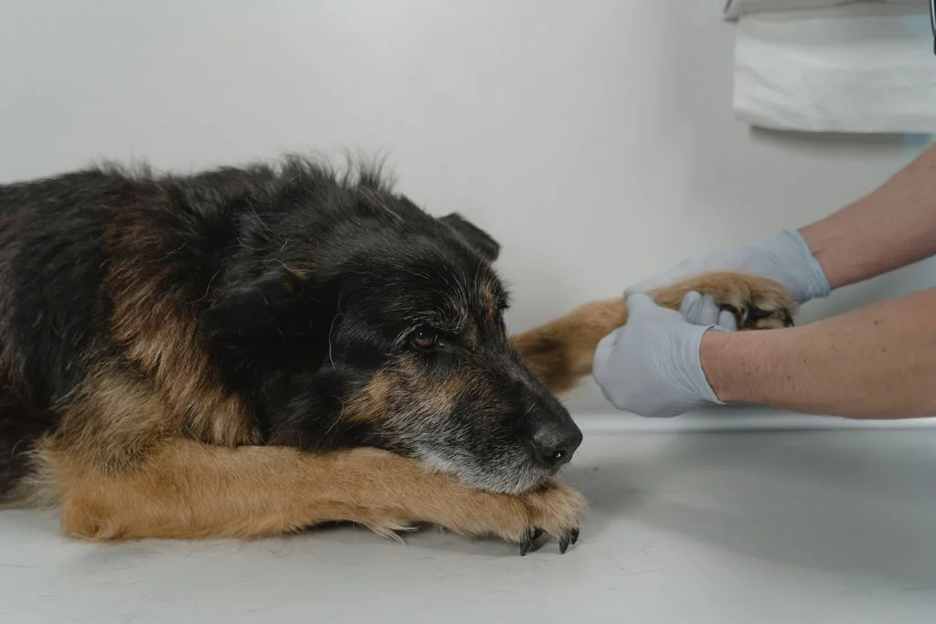
What does the removal of a skin tumor look like in dogs and cats?
First, the doctor examines the lump and takes cells from it with a thin needle (fine needle aspiration, cytology). The result allows us to plan how wide the removal needs to be.
The procedure is performed under anesthesia, using a scalpel or CO₂ laser. The removed tissue is always sent for histopathological examination, which provides the final diagnosis.
Did you see a lump? Don’t wait – have it examined
1 cm
A lump the size of a pea or larger
1 month
Has been present for a month or longer
= biopsy
Time for cytological examination
Lumps detected when they are small can usually be removed in one procedure. Observing them “by eye” only gives them time to grow.
Don’t wait if the lump
- grows quickly over a few weeks,
- changes color, shape, or hardness,
- bleeds, has ulcerated, or is oozing,
- itches, and your pet licks or scratches it,
- varies in size,
- appears in a cat – skin changes in cats are more often malignant.
From lump to diagnosis – step by step
Each stage is determined by the veterinarian after examination. Thanks to diagnostics before the procedure, we know what we are removing and how wide.
1
Examination and measurement
We assess the size, location, hardness, and mobility of the lesion as well as the lymph nodes.
2
Fine needle biopsy
A quick, usually painless collection of cells with a thin needle and evaluation under a microscope.
3
Surgical plan
We select the margin and method: scalpel or CO₂ laser. If a malignant change is suspected, we perform X-rays or ultrasound.
4
Procedure
Under anesthesia with monitoring and pain management. Usually discharged home the same day.
5
Histopathology
Final diagnosis and assessment of margins. We discuss the results and the further plan.
Most Common Tumors and Skin Changes in Dogs and Cats
Select to learn what a change is and how we treat it. Remember: diagnosis is made by a veterinarian after examination.
Lipoma
Lipoma – a tumor made of fatty tissue
What is it
A benign tumor made up of fat cells. Very common in older dogs, especially those who are overweight.
What it usually looks like
Soft, movable lump under the skin, usually painless. Grows slowly.
How we proceed
We confirm the diagnosis with cytology. Small lipomas are often monitored. We remove those that grow quickly, compress, or interfere with movement.
Mast Cell Tumor
Mastocytoma – one of the most common skin tumors in dogs
What is it
A tumor that can behave benignly or very aggressively. The degree of malignancy is determined by histopathological examination.
What it usually looks like
It can look like an innocent pimple, wart, or lipoma. Sometimes it changes size from day to day.
How we proceed
We diagnose it with cytology before the procedure to plan a wide margin. We also use medications to protect against the effects of histamine release.
Soft Tissue Sarcoma
Soft tissue sarcoma
What is it
A malignant tumor of the tissues under the skin. Rarely metastasizes, but invades the surrounding area and easily regrows.
What it usually looks like
A hard or soft lump, often mistaken for a lipoma. Usually painless.
How we proceed
A wide margin is crucial during the first procedure. For difficult locations, we plan treatment together with an oncologist.
Papillomas and adenomas
Warts, sebaceous adenomas – “cauliflowers” in older dogs
What it is
Benign changes of the skin or sebaceous glands. There are often several of them.
What it usually looks like
Small, pink or gray growths with an uneven surface, sometimes bleeding when scratched.
How we proceed
Ideal for CO₂ laser removal – precisely, with a small wound, even several changes during one procedure.
Histiocytoma
A lump typical for young dogs
What it is
A benign skin change, most often in dogs under 3 years of age. It often resolves on its own within a few weeks.
What it usually looks like
A rapidly growing, pink, hairless bump in the shape of a button, often on the head, ears, or paws.
How we proceed
We confirm the diagnosis with cytology. Usually, we observe it. We remove it if it does not resolve, ulcerates, or causes discomfort.
Cysts and lipomas
Epidermoid cysts, blocked glands
What it is
Fluid-filled vesicles in the skin. They can burst, become inflamed, and recur.
What it usually looks like
Round, well-defined lumps, sometimes with an opening from which a grayish mass emerges.
How we proceed
We remove the cyst completely, along with the sac, often using a CO₂ laser. Do not squeeze it at home – it risks inflammation.
Why the first operation is the most important
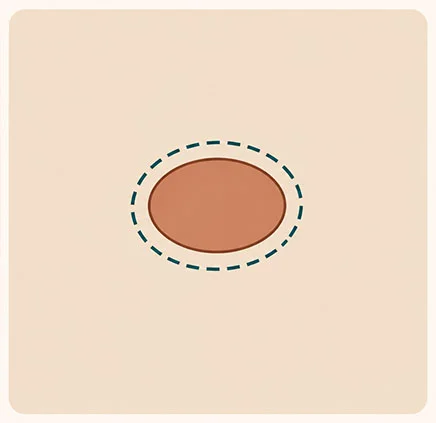
Illustrative diagram. Created by AI.
Benign change
A benign change, such as a lipoma or cyst, can be removed with a narrow margin of healthy tissue.
The wound is small, and healing is quick.
That is why we do not remove lumps “blindly”. First, a diagnosis, then a procedure planned to remove the change completely the first time.
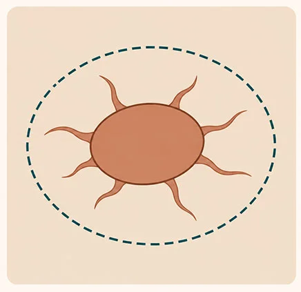
Illustrative diagram. Created by AI.
Malicious Change
Malignant tumors send out invisible “tentacles” into the surrounding tissue. To completely remove it, it must be excised with a wide margin, even 2–3 cm of healthy tissue.
The first surgery offers the greatest chance of cure – each subsequent one is more difficult.
That is why we do not remove lumps “blindly”. First, a diagnosis, then a procedure planned to remove the change completely the first time.
CO₂ Laser Tumor Removal – for Small and Large Lesions
With CO₂ laser, we remove both small and large skin tumors, even with a wide margin when the type of lesion requires it. When the tumor is located in an area where the margin must be narrow – e.g., on a finger or eyelid – the surgeon may use a laser or scalpel, or combine both methods, so that the edges of the removed tissue are fully clear for the histopathologist. The laser simultaneously cuts and seals small vessels, resulting in less bleeding and swelling. Laser procedures are performed by Dr. Małgorzata Szczepańska, trained in laser surgery. The choice of method is determined by the surgeon.
More about laser surgery →
Less bleeding
Small vessels close during cutting, so the surgical field is clean.
Smaller swelling
Closed lymphatic vessels reduce swelling after the procedure.
Small and large changes
Laser works well for both minor changes and large tumors requiring a wide margin.
Precision in difficult places
Eyelids, lips, fingertips, or spaces between fingers – the laser allows for very precise work.
What you should know about skin tumors
- Most skin nodules in dogs are benign, but some turn out to be malignant. Even an experienced veterinarian cannot distinguish them by appearance alone.
- Not every nodule needs to be removed immediately. Small, confirmed lipomas are often just monitored. However, every nodule requires evaluation.
- If the histopathological examination shows a malignant change or an incomplete margin, a second procedure or an oncology consultation may be needed.
Care after tumor removal
Collar or postoperative garment
Protects the wound from licking until healed, usually about 10–14 days.
Check the wound twice a day
Mild swelling and redness in the first days are normal. Do not apply anything to the wound without recommendation.
Limited movement
Walks on a leash, no running, jumping, or bathing until the wound is healed.
Histopathology result
We will discuss it with you during the follow-up visit or by phone along with the further plan.
Call immediately if:
- the wound is bleeding, there is pus oozing from it, or it has opened up,
- a large, hard swelling has appeared at the site of the procedure,
- your pet is not eating, is lethargic, or has a fever,
- vomiting or diarrhea has occurred after the removal of a mast cell tumor.
Removal of skin tumors at Sosken


Questions about skin tumors
Did you find a lump on your pet? Schedule an examination – a quick cytology often explains a lot.
FAQ – questions and answers
Based on the studies
Graf R. et al. (2018), Veterinary Pathology
Skin tumors in dogs – data from the Swiss canine cancer registry from 2008–2013: what changes occur most frequently and in which breeds. – doi: 10.1177/0300985818789466 →
Kiupel M. et al. (2011), Veterinary Pathology
Two-step histopathological grading system for mast cell tumors in dogs – why histopathological examination is crucial for prognosis. – doi: 10.1177/0300985810386469 →
A lump is a question. The answer is provided by examination
Schedule an appointment. We will check the change, and if necessary, we will plan a procedure – also with CO₂ laser.